 Isaiminia World Breaking News & Top Stories
A fetal echocardiography is a test that enables your doctor to see the heart of your developing child. This ultrasound examination can find a heart defect before your baby is born by using sound waves to look at the heart’s architecture. The heart of your unborn child is depicted in greater depth in a fetal echocardiography than in a standard prenatal ultrasound. If you’re looking for an ultrasound center near you to schedule a fetal echocardiography, search for ultrasound center near me online or consult with your doctor for recommendations.
What is fetal echocardiography?
An essential prenatal examination that reveals the details of your unborn child’s heart is a fetal echocardiography. This test is carried out by medical professionals to look at the heart’s rhythm and blood flow. A fetal echocardiography can be used to determine how well a baby’s heart is pumping.
Reasons to have an echocardiogram:
A fetal echocardiography is required in a few circumstances during pregnancy. Have a fetal echocardiography for the following reasons:
- Family history of heart illness or congenital heart abnormalities
- During a routine ultrasound, an abnormal heart rhythm or another heart issue may be discovered.
- Diabetes, lupus, or phenylketonuria in the mother
- During the first trimester, the mother’s rubella test was positive.
- Medicine use by mothers that could harm a baby’s developing heart.
- Chromosome abnormality that was identified after an amniocentesis.
- Pregnancy after in vitro fertilization (IVF)
- Monochorionic twins (two unborn children who share a placenta)
When should you undergo fetal echocardiography scan?
It is best to get a fetal echocardiogram between weeks 18 and 22 of pregnancy. Parents will feel more at ease if the fetal echocardiography is normal. If a serious heart condition is found during this test, prospective parents are given advice regarding the condition, including what to expect at birth, what preparations need to be made, what types of procedures will be necessary and at what ages, what the long-term outlook is following repair, etc. It is possible to inform and counsel parents about the disease so they may decide on an appropriate course of treatment.
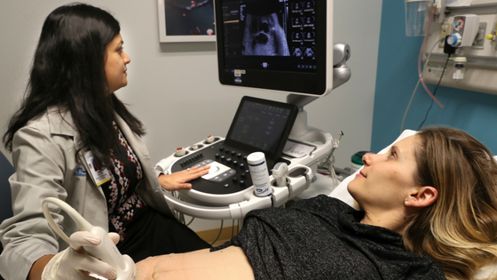
Procedure:
A fetal echocardiography is most frequently performed using abdominal ultrasound. To take images of your baby’s heart, the healthcare professional must place the ultrasound probe over your tummy. Your doctor will begin by rubbing a warm gel onto your stomach. The image displayed on the monitor is the result of sound waves being emitted by the ultrasound probe and returning from your baby’s heart. An endovaginally ultrasound can also be used to perform a fetal echocardiography. A tiny ultrasound transducer is inserted into the vagina during this kind of test to take images of your unborn child’s heart. Early in pregnancy, a transvaginal ultrasound can be done and gives a sharper picture. A registered cardiac sonographer (RCS), a medical professional with specialized training in using ultrasound equipment and scanning a baby’s heart, typically performs a fetal echocardiogram. A pediatric cardiologist examines the test findings when the surgery is complete. This physician has received specialized training in identifying and treating cardiac problems in infants and young children.
Result of fetal echocardiography:
A fetal echocardiography is performed to look for cardiac issues in your unborn child. These issues could be caused by the way the heart develops or functions. Arrhythmias, or irregularities in the baby’s cardiac rhythm, may also be visible. Usually, a fetal echocardiography is repeated to confirm the findings when it shows that your baby’s heart has a problem. Following that, you will frequently have meetings with the pediatric cardiologist to discuss the heart problem and how it can influence your baby’s general health.
Complications related to fetal echocardiography tests:
There is no proof that FE done at a gestational age of more than 18 weeks (about 4 months) is hazardous to either the mother or the fetus. FE is not painful. For some newborns, early heart problem discovery could mean life or death. Additionally, it might help prevent issues like high pressure in the lung veins, which could render the child incapable of being operated on. If the baby is known in advance, all preparations can be made for it.
Summary:
An ultrasound test called fetal echocardiography is used to look at the developing baby’s heart’s structural components. This examination can find heart issues like faults or arrhythmias. If you have a family history of congenital heart abnormalities, your doctor could advise getting a fetal echocardiography. It could take anywhere from 45 minutes to two hours to complete the process. Usually, results are available in 24 hours.