 Isaiminia World Breaking News & Top Stories
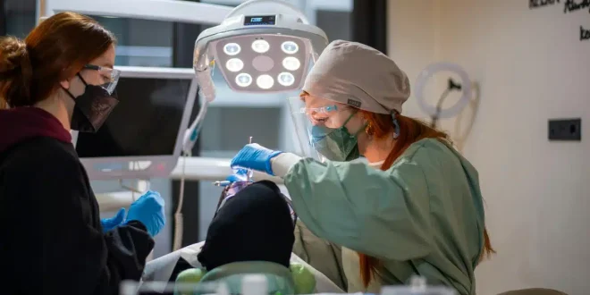
Your teeth often stay silent when problems start. Small cavities, gum disease, even early signs of oral cancer can grow without pain. General dentistry now uses imaging tools to catch these threats early. You see more than a quick glance in a mirror. You see clear pictures that reveal what your eyes cannot. X‑rays, 3D scans, and digital photos help your dentist spot tiny changes before they turn into deep damage. As a result, treatment stays simple. Recovery stays shorter. Costs stay lower. The same technology that plans complex work like Chelsea dental implants also protects natural teeth. It shows bone loss, hidden decay, and infections long before they spread. Early detection is not a luxury. It is basic protection for your health, your comfort, and your ability to eat and speak without fear.
Why early detection matters for you and your family
Tooth decay and gum disease grow in stages. You often feel nothing during the first stage. By the time you feel pain, damage has already spread.
Early detection helps you
- Keep more of your natural teeth
- Need less drilling and fewer shots
- Spend less time in the chair and at home recovering
Silent problems also affect your whole body. Untreated gum disease links with heart disease and diabetes. The Centers for Disease Control and Prevention explains that strong oral health supports your general health. When imaging tools catch trouble early, you protect more than your smile.
Main imaging tools your dentist may use
General dentistry today uses three common types of imaging.
- X ray images
- 3D cone beam scans
- Digital photos and intraoral camera images
Each tool shows a different part of the story. Together, they give a clear view of teeth, gums, bone, and soft tissue.
How X rays support early detection
Standard X-rays remain basic tools in every office. They show what lies between teeth and under fillings where eyes cannot see.
With X rays your dentist can
- Find small cavities between teeth
- See infections at the tip of roots
- Measure bone loss from gum disease
Modern digital X-rays use less radiation than older film systems. The U.S. Food and Drug Administration states that dental X-ray exposure stays low and safe when used only when needed. You can ask how often you need them based on your risk and history.
How 3D scans change complex care
Some offices use cone beam computed tomography. This tool creates a 3D view of your teeth, jaw, and nearby structures.
3D scans help when you
- Need dental implants
- Have impacted teeth
- Show signs of cysts or tumors in the jaw
The same 3D view that guides safe implant placement also helps spot hidden disease. Your dentist can see narrow cracks, deep infections, and bone changes that a flat X-ray might miss. Early action often means a simple filling instead of a root canal or extraction.
How digital photos and cameras help you see
Digital cameras and small intraoral cameras show real-time images on a screen. You see what your dentist sees.
These tools help with early detection because they
- Show early white spots that signal the start of decay
- Reveal small chips and wear from grinding
- Record changes in gums and soft tissue over time
Visual proof builds trust. You do not need to imagine a problem. You see it. That makes it easier to agree to small steps now instead of large treatment later.
Comparing common dental imaging tools
| Imaging tool | What it shows | Best for early detection of | Typical use in general dentistry
|
|---|---|---|---|
| Bitewing X rays | Crowns of upper and lower teeth | Small cavities between teeth and early bone loss | Routine checkups every 1 to 2 years based on risk |
| Periapical X rays | Whole tooth from crown to root tip | Infections at root tips and deep decay | When you have pain or deep fillings |
| Panoramic X ray | Full jaws, sinuses, and joints | Impacted teeth, cysts, and jaw issues | Planning braces, extractions, and implants |
| Cone beam 3D scan | 3D view of teeth, bone, and nerve paths | Hidden fractures, bone defects, and implant planning | Complex cases and surgery planning |
| Digital photos and intraoral camera | Surface view of teeth and gums | Early enamel changes and gum changes | Baseline records and patient education |
Safety and common worries about imaging
Many people fear radiation. That fear is natural. It also often comes from old stories.
Today imaging uses
- Low dose digital sensors
- Fast exposure times
- Lead aprons and thyroid collars when needed
Your background radiation from daily life stays higher than your yearly dental X-rays in most cases. You can still ask if each image is needed. A dentist who respects your health will explain the reason for each test in clear terms.
How imaging protects children, adults, and older adults
Every age group gains specific benefits from early detection.
Children
- Monitoring growing jaws and incoming teeth
- Finding weak spots before cavities form
- Planning space for adult teeth
Adults
- Catching stress cracks and wear from grinding
- Tracking gum health and bone levels
- Planning crowns and implants with less risk
Older adults
- Watching bone levels around implants and bridges
- Finding root decay near the gum line
- Checking fit of dentures and partial dentures
How you can support early detection
You play a key role in early detection. Imaging works best when you combine it with steady habits.
Three key steps help
- Keep regular checkups and cleanings
- Follow your dentist’s schedule for X rays and photos
- Speak up about any change in your mouth
Even small signs matter. A sore that does not heal. A tooth that feels “off”. A change in how your teeth meet. When you speak up, imaging can focus on the right spot and give quick answers.
Moving from fear to control
Many people avoid the dentist out of fear. They fear pain, cost, or bad news. That delay often leads to larger problems.
Imaging tools offer control. They show problems when they are still small, quiet, and easy to fix. They turn guesswork into clear facts. With those facts, you and your dentist can plan simple, timely care.
Your mouth is part of your body. Protect it with the same care you give your heart or your eyes. Use imaging as a shield, not a threat. Early detection gives you more choices, more comfort, and more years with your own teeth.